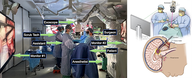
Optimal intraoperative visualization has been the cornerstone for the safe, successful completion of brain tumor removal. The recent advent of the digital surgical exoscope has provided a high definition and magnified three-dimensional (3D) panoramic view on a heads-up display that can be used for performing surgery. The exoscope provides a long-working distance, high magnification, and field depth to permit neurosurgery without surgeon obstruction. LED illumination by the exoscope permits reduction in thermal damage and tissue glare that can occur with the conventional microscope.
The exoscope camera system is positioned alongside the neurosurgeon in a hands-free manner with use of a robotic independent arm to permit delineation of tumors in the brain. Surgeon ergonomics are improved with use of the exoscope promoting a more relaxed posture and alleviating neck and back strain. The surgeon remains upright in a neutral position, minimizing back or neck tilt and minimizing strain on the neck or back.
The exoscope can also be used to perform fluorescence-guided surgery (FGS) with appropriate light filters that include 5-aminolevulinic acid (5-ALA) for glioblastoma (GBM) surgery. In addition to using the exoscope to visualize tumors, important tractography is integrated with the exoscope neuronavigation system to identify and avoid important tracts that are involved with movement, speech, and vision.
Costas Hadjipanayis, MD, and PhD, and his team at UPMC routinely use the robotic-assisted exoscope for all brain tumor surgeries. His team was one of the first to report optimal patient outcomes for brain tumor patients undergoing removal of their GBM, brain metastasis, pineal, and posterior fossa tumors with use of the exoscope.

The exoscope provides a unique advance for the surgical treatment of brain tumors. For patient enquiries please call 412-647-0953.
